
Angina refers to chest pain caused by reduced blood supply to your heart. Those experiencing an episode of angina may confuse it with a heart attack. However, a heart attack occurs when the blood supply to your heart is suddenly blocked, which is life-threatening. Angina, on the other hand, is not immediately life-threatening but is a sign that the arteries that supply your heart (coronary arteries) are not healthy.
Is your chest pain angina?
Chest pain that is sharp, changes with breathing or movement and/or is accompanied by tenderness on touch is unlikely to be angina.
In contrast, chest pain that feels crushing and occurs in the centre of your chest is likely due to an attack of angina or a heart attack.
Chest pain caused by a heart attack is sudden, severe and usually accompanied by other symptoms, including sweating, nausea and vomiting.
Chest pain caused by angina is a long-term (chronic) condition. It occurs frequently on exertion when the oxygen demands to your heart goes up with your heart rate consequently increasing. It goes away with rest or in response to a glyceryl trinitrate (GTN) spray. To understand why, it helps to first understand the anatomy of your heart.
The anatomy of your heart
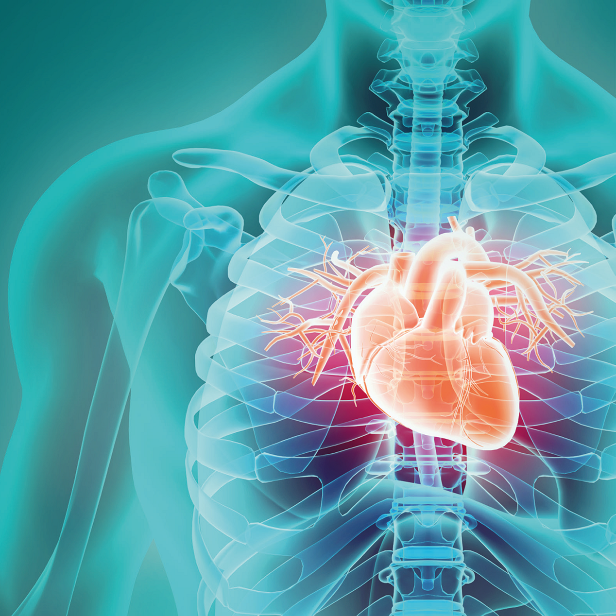
Your heart is a powerful pump containing four chambers: two upper chambers called atria and two lower chambers called ventricles. The activity of your heart is controlled by electrical impulses that spread from the upper chambers to the lower chambers. This allows your heart to pump oxygen-rich blood throughout your body, as well as to supply its own tissue with oxygen-rich blood.
There are three main blood vessels that supply your heart tissue with oxygen-rich blood. These are called coronary arteries, specifically the right coronary artery and the two branches of the left coronary artery, the left anterior descending artery and the circumflex artery.
Each artery is made of three layers; the inner layer or endothelium (tunica intima), the middle layer of smooth muscle (tunica media) and the outer protective layer (tunica externa).
The inside of healthy coronary arteries is smooth with no blockages, allowing for the easy, smooth flow of blood known as laminar flow.
Unhealthy coronary arteries
Coronary arteries that are diseased are not smooth on the inside and instead are lined with fatty deposits (atherosclerotic plaques) that restrict the flow of blood.
The risk of your coronary arteries becoming blocked by plaques increases with age, certain health conditions (eg diabetes, high blood pressure and high cholesterol), lifestyle factors (eg smoking, carrying excess weight) and a family history of coronary artery disease. Your risk is also higher if you’re male and/or of South Asian descent.
As the size of these plaques increases, the space with an artery (lumen) reduces. Coronary artery disease occurs when that space is reduced by around 30%. At this point, you’re unlikely to have any symptoms. When this space is reduced by around 70–80%, angina develops.
Once plaques form, there is the risk that they can break off (rupture) due to aggravating factors, such as inflammation. This can cause platelets to build up and form a clot (thrombus), which can completely block a coronary artery — this causes a heart attack.
Angina symptoms and types
Angina can be broadly grouped into two types: typical and atypical.
Typical angina meets three specific criteria: it occurs on exertion (eg when climbing the stairs, running or exercising) or during emotional stress, and it is relieved by rest or a GTN spray, which widens (dilates) the arteries.
Atypical angina only meets two of these three criteria. For example, when chest pain occurs only at rest.
If you experience any of the above symptoms of angina, it’s important to see a doctor to have your symptoms investigated.
Diagnosing angina
Getting a diagnosis of angina depends on three factors: your symptoms, your medical history and a clinical examination, which may involve specific tests.
If you experience chest pain on exertion that goes away with rest or with a GTN spray, you most likely have angina. If you don’t meet all of these criteria, you may still have coronary artery disease and angina.
Signs of obstructive coronary artery disease, where your coronary arteries are gradually narrowing, can include a rapid heart rate (tachycardia) or raised jugular venous pressure, which can be determined by examining your neck.
To confirm a diagnosis of angina, you will likely need to undergo tests. In the past, this usually involved bringing on your suspected angina symptoms by doing exercise (eg on a treadmill or stationary bicycle) while closely monitored. However, modern testing techniques are now more effective and sensitive.
Angina tests
Today, testing for suspected typical angina involves a coronary angiogram.
During a coronary angiogram, a thin tube (catheter) is inserted into the radial artery of your arm. Using X-ray imaging, the catheter is guided to your heart. A dye is injected through the catheter to highlight your coronary arteries. X-ray images are captured to reveal if any of your coronary arteries are blocked and to what extent.
A coronary angiogram is a low-risk, minimally invasive procedure. Nonetheless, if you have atypical angina, your doctor will likely recommend a less invasive test to image your coronary arteries, namely a cardiac CT scan, also known as a CT coronary angiogram.
Other non-invasive tests include stress testing where you take medication to increase your heart rate, which consequently increases the amount of oxygen your heart needs, causing it to work harder.
While your heart is under stress, your doctor may perform an electrocardiogram (ECG), echocardiogram or MRI scan to detect and measure signs of any blockage. If you have a blockage in your coronary arteries, you will develop angina symptoms associated with changes in your ECG or MRI scan.
Heart treatments
Diagnosis and specialist treatment for a wide range of heart and circulatory conditions.
Treating angina
It is important to treat angina to reduce your risk of a heart attack and prolong your life (ie improve your prognosis), as well as to ease your symptoms.
Treatment involves increasing blood flow to your heart and/or reducing the oxygen demand of your heart tissue.
Most individuals can effectively manage their angina with tablets. To increase blood flow to your heart, your doctor may prescribe tablets, such as nitrates, calcium channel blockers or newer drugs such as ranolazine or nicorandil, which relax the smooth muscle in your coronary arteries. This causes them to dilate, increasing their diameter and letting more blood flow through.
To reduce how much oxygen your heart needs, your doctor may prescribe tablets such as beta-blockers (eg bisoprolol), which reduce your heart rate. With a lower heart rate, your heart doesn’t have to work so hard and therefore doesn’t need as much oxygen.
If medications aren’t enough to resolve your symptoms, your doctor may recommend a coronary angioplasty.
This involves passing a catheter through a vein in your arm and using X-ray imaging to guide it to your heart and into your narrowed coronary artery. A balloon and stent are passed along the catheter. The balloon is inflated to widen the narrowed coronary artery and a stent is left in place to keep it that way after the balloon is deflated and removed.
Once you’re undergoing treatment for angina, it is still helpful to make lifestyle changes, such as losing excess weight, quitting smoking, exercising regularly and following a healthy, balanced diet to reduce high cholesterol.
