
MRI stands for magnetic resonance imaging and refers to the use of powerful magnets and radio waves to create 2D and 3D images of your bones and soft tissues. MRI scans are used to capture detailed images of what’s going on within your body and unlike an X-ray or CT scan, do not use radiation.
Why would you need an MRI scan?
There are lots of different reasons why a doctor may recommend you have an MRI scan, all of which relate to checking the health of your internal tissues, which include your bones, soft tissues (such as blood vessels, ligaments and muscles) and the brain. Sometimes an MRI scan may be used to diagnose an injury or disease, while at other times it may be used to check your progress in response to treatment you’re receiving.
Common parts of the body an MRI scan may be used to investigate include:
- The brain and spinal cord — to detect the extent of brain damage caused by stroke, scars to the brain and spinal cord caused by multiple sclerosis, the development of tumours and many other neurological conditions
- The heart and blood vessels — to detect heart disease, damage caused by heart attacks, as well as structural problems with the valves of the heart and blood vessels
- The bones and joints — to detect injuries and damage, as well as bone infections and cancer
MRI scans are also used to investigate the health of other organs, including the bladder, bowel, liver, kidneys and pancreas. They can also help evaluate the health of the ovaries and breasts in women and the prostate in men, usually in the context of detecting cancer.
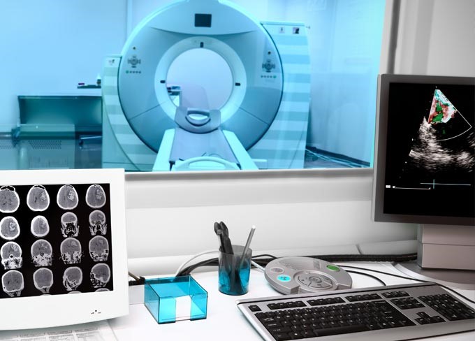
Depending on the reason for your MRI scan, you may have a special dye injected into your body to help certain tissues show up more clearly. The scan itself involves lying down in a large tube which contains powerful magnets; this is an MRI machine.
How does an MRI machine work?
The technology behind MRI machines has been around since the early 1970s. It began with American chemist, Paul Lauterbur, who developed the process and named it zeugmatography. He went on to win the Nobel Prize for this ground-breaking work. By the 1980s, MRI scanning was used in hospitals across the UK to help diagnose a range of medical conditions.
MRI machines today work on the same principles as they did over 40 years ago, namely the creation of strong magnetic fields that affect the water molecules in the body. As on average 60% of the human body is made of water, according to the NHS, MRI scanning can capture a detailed picture of the body’s internal tissues.
The physics of MRI scanning
Every water molecule contains two hydrogen atoms, and within each hydrogen atom is an even smaller particle called a proton. Protons act like microscopic magnets, which makes them sensitive to magnetic fields. A strong enough magnetic field, such as that produced by the large, powerful magnets of an MRI machine, causes these protons to line up in the same way.
Next come the radio waves. Bursts of radio waves excite the hydrogen atoms in the body, which forces the protons within them out of alignment. When the radio waves stop, the protons realign themselves. This releases radio signals which can be picked up by sensitive detectors built into the MRI machine. Protons in different tissues realign at different speeds, which is why different types of tissue appear differently on an MRI scan.
The radio signals detected from the protons as they realign are sent to a computer where they are used to create images of the body’s tissues. The computer sits in a separate room to the MRI machine as the machine’s magnets are so powerful that they interfere with computers.
No metals allowed
The strength of MRI machine magnets also means that no metal items (such as glasses, earrings, watches and zips) should be worn during a scan. Some, but not all, metal implants, such as pacemakers and artificial joints, may prevent you from having an MRI as they can malfunction or interfere with the imaging process. Modern pacemakers and artificial joints are usually MRI-compatible but in either case, you should let your doctor know about any metal implants you have.
Are there different types of MRI scan?
The short answer is yes. Different types of MRI include functional MRI, cardiac MRI, breast MRI, MRA (magnetic resonance angiography) and MRV (magnetic resonance venography). They are each used to investigate different tissues.
Functional MRI
Functional MRI focuses on imaging the brain. Where a standard MRI scan of the brain highlights the structures within it, a functional MRI scan looks at brain activity when carrying out different tasks, such as talking, recalling memories or moving. This is possible because more active parts of the brain show an increase in the flow of blood carrying oxygen. Functional MRI detects this change in oxygen levels.
Functional MRI is used to help plan brain surgery and determine the effects of brain trauma and conditions such as stroke or Alzheimer’s disease.
Cardiac MRI
Cardiac MRI focuses on imaging the heart to detect heart disease or other heart problems. It can reveal both the structure of the heart as well as how well it is working, for example, by looking at how well blood flows through the heart.
Breast MRI
A breast MRI scan is sometimes used to screen for breast cancer in younger women at high risk of the disease, for example, if they have a strong family history of breast cancer. It is also used to determine the extent of breast cancer in women who have already been diagnosed.
MRA
MRA focuses on imaging blood vessels, specifically the arteries. A special dye is injected before the scan to help visualise the blood vessels more clearly. This type of MRI scan is used to examine the structure of blood vessels to detect problems such as narrowing or blockage.
MRV
MRV also focuses on imaging blood vessels, specifically the veins. As with MRA, a special dye is injected to make it easier to image the veins. This type of MRI scan is used less frequently than MRA, but is useful for detecting structural problems with the veins and diagnosing conditions where blood clots occur in the veins, such as deep vein thrombosis and cerebral venous thrombosis.
What do the results of an MRI scan look like?
Whichever the type of MRI scan, the resulting images are always black and white, with each image representing a virtual slice through the body. Black parts of the image represent air or hard bone, while soft tissues and fluids appear different shades of grey or white. Your doctor will look for differences in shades and contrast to identify the different tissues and whether or not a tissue has been injured or is diseased.
More about MRI scans
Your doctor may recommend an MRI scan for a number of reasons, including joint pain, heart health, bowel conditions and more. If you would like to learn more about having a private MRI scan at a Spire hospital, please visit our Spire Healthcare MRI page.
