
Spire Healthcare’s Shawfair Park and Murrayfield Hospitals in Edinburgh both secure ‘Good’ outcomes
27 November 2024
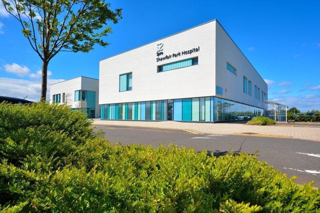
Spire Shawfair Park Hospital, part of Spire Edinburgh Hospitals, has received another ‘Good’ outcome from an unannounced inspection by Healthcare Improvement Scotland (HIS) in September. This complements a similar ‘Good’ outcome awarded to Spire Murrayfield Hospital following a previous unannounced HIS inspection visit, in August.