
The sooner you’re better, the better
Spire London East Hospital
See an expert in as little as 48 hours

The sooner you’re better, the better
Spire London East Hospital is a well-established private hospital delivering comprehensive treatments and care to our patients in east and central London and Essex. We offer fast access to high-quality healthcare, from consultations and advanced diagnostics to personalised treatments and expert aftercare.

How can we help?
Search or browse by treatments or consultants to see what Spire can do for you.
Treatments
Looking for a specific treatment or procedure?
Consultants
You can expect outstanding care from our expert consultants and dedicated nurses.
Events
We have a regular programme of events happening in and around our hospital.
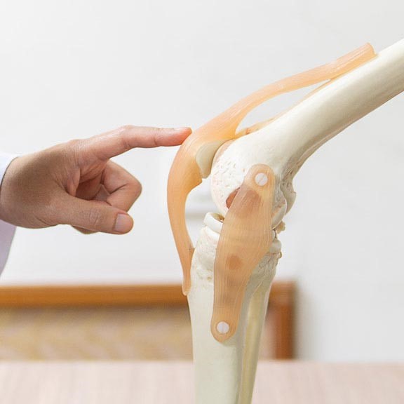
Bones and joints
Through surgery or less invasive procedures like physiotherapy, our team of orthopaedic specialists may be able to help ease the pain in your bones and joints to get you back to living the life you love.
Spire London East Endoscopy Unit is JAG accredited
Offering fast diagnosis of digestive conditions
The JAG accreditation shows our commitment to patient care through excellent communication and investment in state-of-the-art equipment.

Children and young people services
Specialist clinical care tailored for infants, children and adolescents.

MRI scans
Spire London East Hospital offers a high quality, comfortable and fast MRI scanning service. We offer appointments to fit around you.


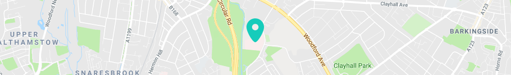
How to get to us
We are located just a couple of miles from the M11 and only 20 minutes on the Central line tube from the city of London. London City Airport is a short car journey of 20 minutes and Stansted airport is just a 30 minute drive.
Spire London East Hospital,
Roding Lane South
Redbridge
Essex
IG4 5PZ
Travel instructions
By tube
Redbridge Underground station is walking distance from the hospital.
By bus
The hopper bus service 366 from Redbridge station stops opposite the hospital.
Parking
We have 152 car parking spaces, which are complimentary to all patients and visitors.
From the M25 junction 27
Follow the M11 towards London. Leave the M11 at junction 4, stay in the left hand lane following signs for A12/Chelmsford. At Redbridge Roundabout take the first exit (next to the Beefeater Restaurant) into Redbridge Lane East. Take the immediate left into Roding Lane South. Spire London East Hospital is one mile along on the left.
From the A406 (North London, Chingford, Walthamstow)
Follow the A406 (North Circular Road) towards London Docklands. Leave the A406 at Redbridge Roundabout slip road (signposted A12/Chelmsford). At the roundabout take the first exit (next to the Beefeater Restaurant) into Redbridge Lane East. Take the immediate left into Roding Lane South. Spire London East Hospital is one mile along on the left.
From the A406 (East London, Barking, Docklands)
From East London, follow the A406 (North Circular Road) towards North London. Leave the A406 at the Redbridge Roundabout slip road (signposted A12/Chelmsford/ Central London). At the roundabout take the third exit (next to the Beefeater Restaurant) into Redbridge Lane East. Take the immediate left into Roding Lane South. Spire London East Hospital is one mile along on the left.
From Gants Hill roundabout (where the underground station is situated, postcode IG2 6UD)
Take the exit marked A1400, Woodford Avenue (towards Walthamstow/Woodford). Continue on to the next roundabout, take the third exit on to the A1400 (signposted South Woodford, Walthamstow and North Circular). After around one mile, turn left on to Roding Lane South (just before the Euro Car Parts warehouse). Continue along Roding Lane South for around a third of a mile, then turn right into Spire London East Hospital.
what3words
///goals.talent.staple
Contact details
Self-pay treatment enquiries
0208 709 7817
PMI treatment enquiries
0208 709 7878
Scans and diagnostics
0208 709 7856
Physiotherapy and sports injuries
0208 709 7779
Invoice and payment queries
0118 975 4118
Hospital Director
Chris Burrows
We record calls to help improve our service. To learn more about how we use your information, read our privacy policy.
Patient reviews
How to book
Whether you’re paying for yourself, using private medical insurance or are an NHS patient, accessing our hospitals is really easy.
Healthcare professionals
A guide for GPs and other healthcare professionals to the services and specialties we offer.